CIRS Model 013 Stereotactic Needle Biopsy Training Phantom提供了一个简单的乳腺摄影针活检程序培训解决方案。
CIRS 013乳腺模体特征:
可压缩
刺破时不会泄漏
包含各种大小密集的活体组织
包含用于可视化和活组织检查的钙化簇
拟人化的形状准确模拟乳房的压缩
模拟针的阻力
物理一致性与人体组织相似
兼容独立和附加立体定位活检系统
适用于数字系统
DESCRIPTION
A tissue equivalent, compressible biopsy training phantom, that wont leak! Features: Compressible Does not leak when punctured Contains various sizes of dense masses for biopsy Contains calcification cluster for visualization and biopsy Anthropomorphic shape for accurate simulation of breast compression Simulated needle resistance Palpable lesions Physical consistency similar to human tissue Compatible with both stand alone and add-on stereotactic biopsy systems Country of Origin: United States of America Stereotactic Needle Biopsy Training Phantom is a disposable training tool and practice medium for mammographic needle biopsy procedures. The phantom also serves as an excellent quality assurance device for stereotactic systems and should be used whenever a new system is installed or repaired to insure accurate needle placement. The phantom can be used to perform the localization accuracy test in the American College of Radiology's stereotactic breast biopsy accreditation program.
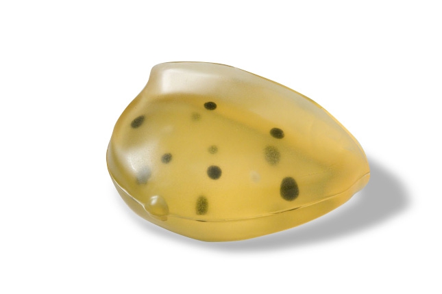
The phantom body is shaped to represent a partially compressed breast. This allows the user to easily compress the breast to 4.5 cm thickness for optimum imaging. The shape also enables the phantom to be used in systems where the patient is either prone or sitting. The phantom is made from a proprietary gel with a physical consistency similar to human tissue and, since the gel is a non-flowing material, it won't leak out when punctured. The gel is surrounded by an elastic skin-like membrane, which enables palpation of the embedded masses and provides needle resistance.
Embedded within the phantom are numerous solid masses in a range of sizes. These radiographically visible masses can be biopsied multiple times. The masses are colored black for easy visualization of a successful biopsy. These solid masses are randomly positioned so each phantom provides a unique training experience. Two calcification clusters are positioned within the medial transverse plane at the right and left edge of the phantom. These calcifications can be biopsied or used for QA checks on the mammography system. To aid in visualizing the location of the calcifications, the two groups are each clustered around a white but radiographically transparent mass.
The stereotactic training phantom offers an easy, low cost option to create a relaxed learning environment. The phantom can be reused multiple times with no special storage requirements. http://szwellcome.b2b168.com